For more than 25 years, Chamberlain Family Orthodontics has been providing patients of all ages with the most effective treatments available. We take pride in the exceptional results we’re able to achieve thanks to the cutting-edge tools we use in our Redlands and Beaumont offices! Digital treatment planning is part of that process, and our patients reap the benefits of this service daily. To learn more about how we make the most of this innovative technology, keep reading below!
3D Scanning
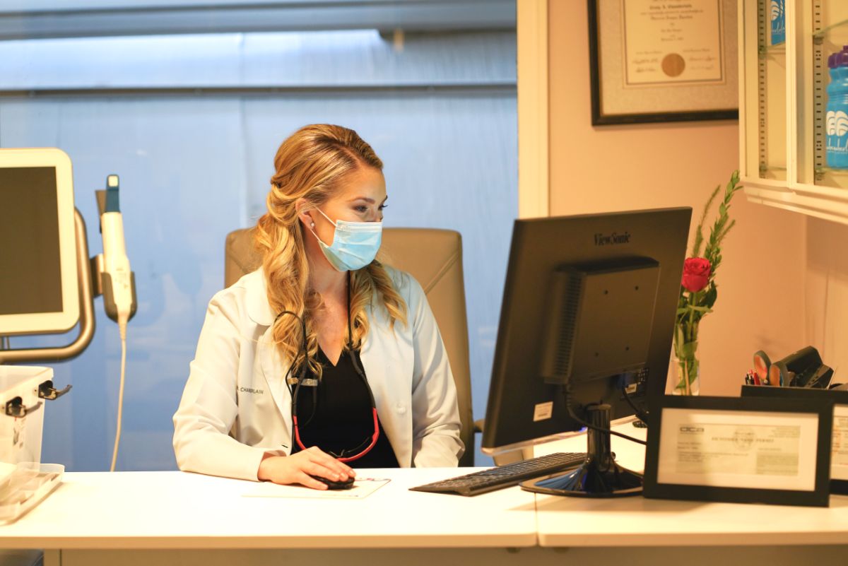
Our practice is always looking for ways to make the orthodontic process less invasive and more efficient for the best results. Adding 3D scanning technology has provided us with an extra element of ease in preparing for treatment! There’s a good deal of prep work involved before active treatment begins, and each step is an important part of the process. These include x-rays, photographs, and a 3D scan of the teeth with our iTero digital scanner.
With the iTero digital scanner, our team uses a small wand to capture a digital image of each tooth and the layout of the gums and mouth. These are saved into our computer system automatically. The scanning process usually takes less than five minutes and provides us with a highly accurate 3D view of the mouth! Once the scan is complete, we can view the images along with the patient and even show them a simulation of the braces or Invisalign treatment plan that’s being considered.
This data is then sent to an orthodontic lab wirelessly so the clear aligners and LightForce brackets can be customized to meet our patients’ smile goals. Everybody involved benefits from a more personalized approach and greater accuracy!

Digital X-Rays
Digital radiographs have become an indispensable tool for helping orthodontists better detect, diagnose, treat, and monitor many oral conditions and diseases. Our x-ray imaging uses digital sensors to replace traditional photographic x-ray film. These can be taken intraorally or extraorally and provide our team with enhanced computer images of the teeth, gums, and other oral structures.
Intraoral x-rays are the most common type of dental x-ray, and can be used to detect cavities, check the status of developing teeth, and monitor teeth and bone health. Extraoral x-rays are able to detect impacted teeth, monitor jaw growth and development, and identify any potential problems between the teeth, jaws, temporomandibular joints, and other facial bones.
Types of intraoral x-rays include the following:
Bitewing x-rays show the upper and lower back teeth in a single view. This type of x-ray is used to evaluate the size of the developing teeth and to check for any signs of decay between the teeth. Bitewing x-rays also help us visualize any bone loss.
Periapical x-rays provide a look at the entire tooth, from the crown to the root and down into the bone that supports the tooth. These x-rays can identify dental problems below the gum line, such as root resorption, abscesses, and bone loss.
Types of extraoral x-rays include the following:
Panoramic x-rays show the entire mouth in one image and are routinely used in orthodontics. They’re also used to plan treatment for dental implants, detect impacted teeth, and identify other jaw problems.
Cephalometric projections show the entire head and help us examine teeth in relation to a patient’s jaw and profile. Our doctors perform precise measurements on these x-rays to evaluate jaw growth and develop an orthodontic treatment plan for a patient.
Digital dental x-rays come with many benefits! These include:
- a digital image that is processed and available to be viewed immediately (compared to traditional film which must be developed)
- up to 80% less radiation exposure than conventional x-rays
- enhanced digital images
- environmentally friendly processing
- electronic sharing
Digital Photographs
In orthodontics, we use both extraoral and intraoral photographs in assessing a patient’s smile. Extraoral photographs give us a good look at the profile and facial proportions of the patient. We use these to evaluate their facial balance and symmetry, lip posture, natural smile line, midline, and more. Intraoral photographs provide us with a visual of the patient’s teeth, gums, and oral tissue from several angles. This shows us how healthy the gums are, the bite alignment, dental issues like crowding and spacing, and the size, shape, and color of the teeth.
What are the benefits of digital treatment planning?
Digital treatment planning helps our doctors obtain exceptionally detailed information about a patient’s oral health, which allows them to build a more effective treatment plan and anticipate any potential challenges. This gives patients a smoother, faster, and more rewarding experience overall! Other benefits include:
- lower risk of surprises during treatment
- a detailed view of your dental and facial anatomy
- less radiation exposure compared to traditional x-rays
- an exceptional level of individualized orthodontic treatment
Get a High-Tech Smile with Chamberlain Family Orthodontics
Chamberlain Family Orthodontics is proud to offer you the latest in orthodontic technology! Our digital treatment planning helps eliminate the mess and extra time involved in traditional processing while giving us the most accurate images and best results possible.
Would you like to learn more about how our digital treatment planning can give you a rewarding experience from start to finish? Get in touch today to schedule a FREE consultation with our talented team!